In vivo 発光・蛍光イメージング
In Vivo Imaging System
| 機器名称 | In vivo 発光・蛍光イメージング In Vivo Imaging System |
|---|---|
| メーカー/型式 |
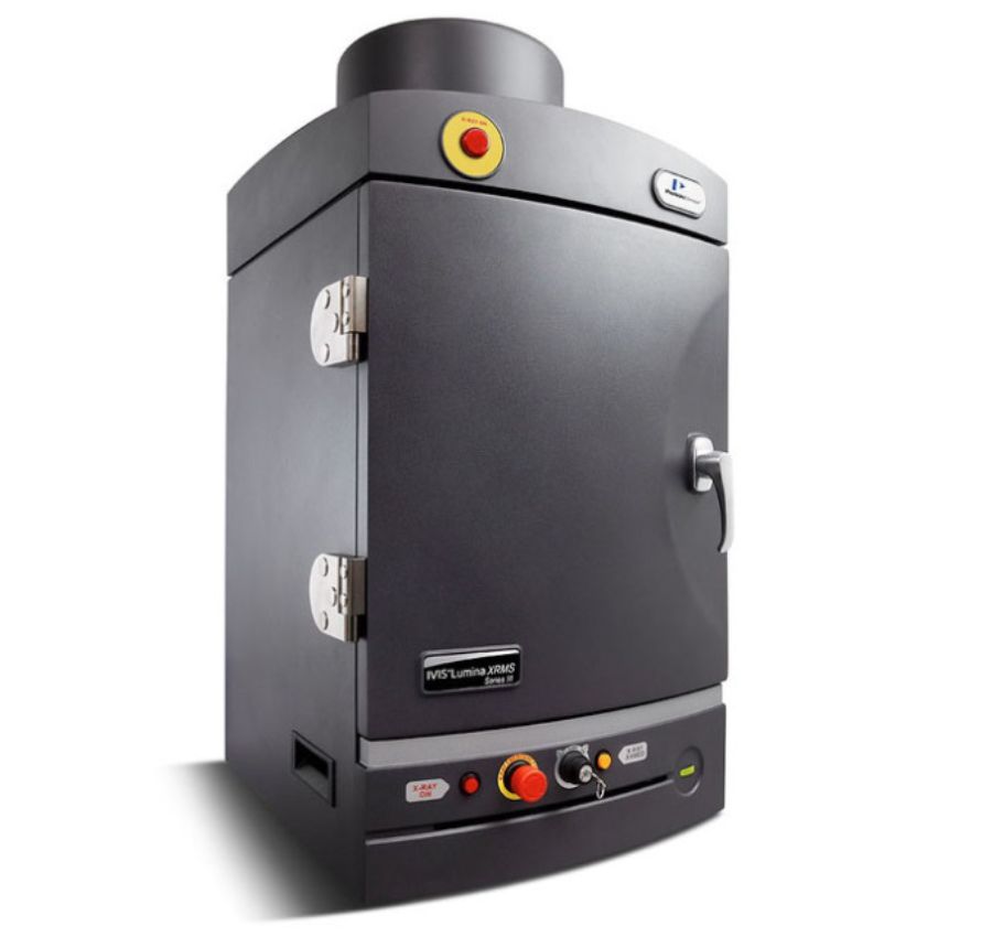
パーキンエルマージャパン/IVIS Lumina XRMS SeriesIII Imaging System |
| 設置場所 |
東大阪キャンパス 39号館N803 |
| 仕様説明 | 本装置は、移植細胞の挙動や薬物動態、更には疾病マーカーをターゲットとした炎症評価等を、非侵襲的に生きたままの生体外から観察(in vivoイメージング)することが可能です。薄型背面照射型ハイグレード冷却CCDと26種類の蛍光フィルタによる高感度光測定能に加え、X線撮影機能も装備されており、発光・蛍光画像にX線写真を重ね合わせることができます。本装置を用いることにより、細菌や癌細胞の増減を光の強度として定量すること、病態モデルなどにおける疾病遺伝子の発現を定量化すること、また、DDSなどを目的とする蛍光イメージング検出することも可能です。更に、豊富な解析ツールの中には6~1536ウェルプレートに対応したROI設定があり、IVISに搭載された超低温冷却CCDカメラを用いた、微弱発光及び蛍光シグナルを数値解析する事が可能です。制御ソフトウェアは、直感的に操作が可能なUIで、初心者に易しい設計となっております。動物倫理にも準じた手軽で簡便な生体画像解析機器として、皆様の多くのご利用を期待しております。 (アプリケーション例:腫瘍学、感染症、移植、薬物動態、炎症など) ー The IVISR Lumina XRMS Series III provides an expandable, sensitive imaging system that is easy to use for two dimensional bioluminescence, fluorescence and X-ray imaging in vivo. The system includes a highly sensitive back-illuminated grade 1 CCD camera, low dose X-Ray imaging system, light-tight imaging chamber and complete automation and analysis capabilities. With the Lumina XRMS, get an anatomical context to optical signal in mice and rats and other large species. The system is equipped with up to 26 filters tunable to image fluorescent sources that emit from green to near-infrared. Furthermore Living ImageR software brings IVIS technology to life by facilitating an intuitive workflow for in vivo optical, X-ray image acquisition, analysis and data organization. The software’s new design creates an intuitive, seamless workflow for researchers of all skill levels. (Key applications in oncology, infectious diseases, implant biology, Inflammation or any model that requires anatomical context) |
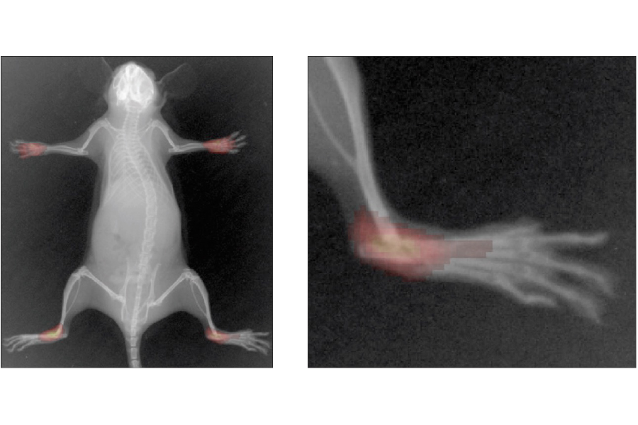
蛍光プローブを用いた関節リウマチの検出。(PerkinElmer社Product Noteより抜粋)
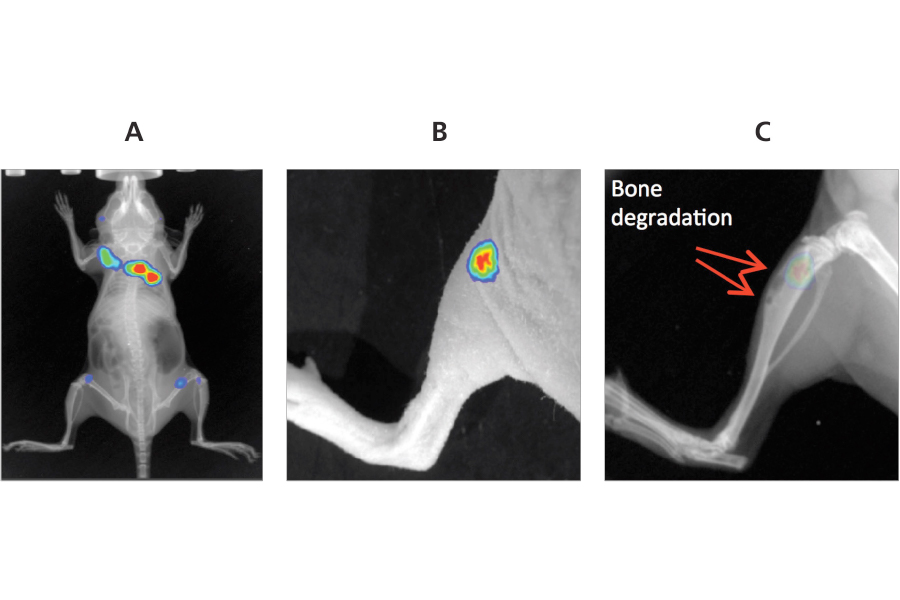
(A)5×10^4個の4T1-luc2細胞を尾静脈注射により静脈内に投与した。 体の様々な部位に定着した細胞を発光で撮影した。(B)発光画像と写真の重ね合わせ。 (C)X線と発光画像の重ね合わせ。 赤矢印は骨溶解領域を示している。(PerkinElmer社Product Noteより抜粋)